Artists have painted cells for more than 350 years

My paintings belong to a genre called science art, or SciArt, that began with a drawing of cork in 1665. In this gallery I gathered the scientists and artists who paint cells, microbes and the molecules inside them, and wrote a title card for every piece.

A young woman in a bright gallery studying a framed painting on the wall

Biology learned what cells look like from drawings

Before cameras could be fitted to microscopes, a drawing was the only way to show anyone else what a cell looked like.

Drawings came first

Robert Hooke's 1665 drawing of cork gave biology the word cell. Santiago Ramón y Cajal's drawings of nerve cells, made for work that shared the 1906 Nobel Prize in Physiology or Medicine, are still in use today.

Scientists still paint

David Goodsell holds a PhD in biochemistry, Greg Dunn a doctorate in neuroscience, and Hunter Cole a PhD in genetics. Each one turns what they know from the lab into art.

Pictures help science stick

In a randomized study of fifth graders, science units taught with the arts built in led to better retention two months later for students reading below grade level.

Drawn at the microscope

The first pictures of cells were made by scientists who looked through a lens and drew what they saw. Their drawings taught the world what a cell looks like.

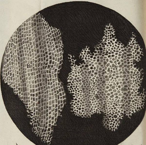
Hooke's drawing of two slices of cork inside a black circle, one showing rows of small boxes and the other a honeycomb of round holes

Cork, from Micrographia

Robert Hooke · 1665, printed plate

Hooke was apprenticed to a painter at 13 before he turned to science, and he drew the plates for his book himself. In a thin slice of cork he saw rows of tiny boxes that reminded him of the small rooms in a monastery, and he called them cells. The word stuck.

Public domain

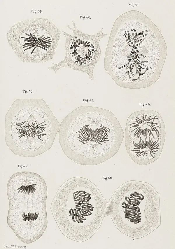
Flemming's drawings of dividing cells, with dark thread-like chromosomes gathering, lining up, pulling apart and finally splitting into two cells

Cell Division, Plate IIIb

Walther Flemming · 1882, printed plate

Flemming drew dividing cells stage by stage, from chromosomes bunched in the middle to a cell pinching in two. In the same book he named the process mitosis, from the Greek word for thread.

Pairs with my Mitosis series.

CC BY 4.0, Wellcome Collection

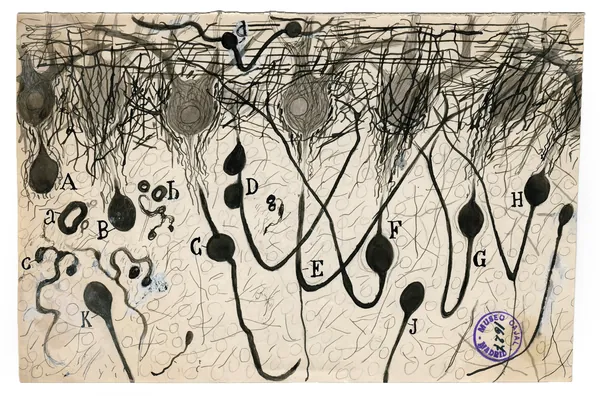
Cajal's drawing of dark nerve cells with long tails and tangled branches on yellowed paper, labeled with letters

Purkinje Neurons

Santiago Ramón y Cajal · about 1900, drawing

Cajal, a Spanish physician and keen painter, stained brain tissue so single nerve cells turned black, then drew them. Purkinje neurons sit in the cerebellum, the part of the brain that coordinates movement. Cajal shared the 1906 Nobel Prize with Camillo Golgi, the scientist the Golgi apparatus is named after.

Pairs with my Golgi Apparatus painting.

Public domain

Painting the inside of a cell

Scientist painters today work from research data and lab images to show what no single microscope picture can: a whole cell, with every part in its place.

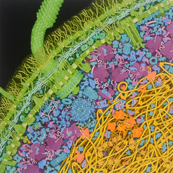
Goodsell's watercolor cross-section of a bacterium: a green cell wall, a yellow tangle of DNA, and crowds of purple, blue and orange molecules

Escherichia coli Bacterium

David S. Goodsell · 2021, watercolor

Goodsell, a biochemist at Scripps Research, paints part of a living cell magnified about a million times, with molecules placed from research data. Here an E. coli cell is cut open: the green wall, the yellow tangle of DNA, and reddish purple ribosomes that build proteins.

Pairs with my Protein Transcription series.

CC BY 4.0, RCSB Protein Data Bank

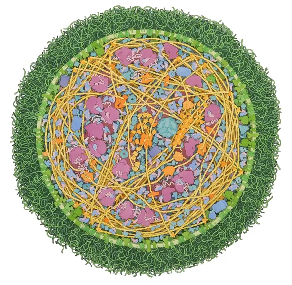
Goodsell's watercolor of a whole round bacterial cell with a fuzzy green edge, packed inside with yellow strands and pink and blue molecules

Mycoplasma mycoides

David S. Goodsell · 2011, watercolor

This painting shows an entire bacterial cell about 300 nanometers across, small enough that a thousand of them in a row would still be thinner than a credit card is thick.

CC BY 4.0, RCSB Protein Data Bank

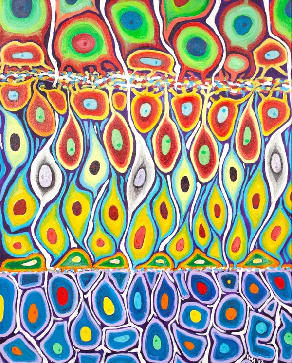
A bright acrylic painting of rows of cells in three layers, each cell outlined in a different color with a colored nucleus

Cell Fates in Zebrafish Retina

Prof. Bill Harris · acrylic painting

An impression of a nearly mature zebrafish retina, its cells settled into three layers and colored by type. The painting was inspired by the multicolored fluorescent proteins researchers use to label different kinds of cells.

CC BY 4.0, Wellcome Collection

Cells as fine art

Some painters treat cells the way other artists treat landscapes and portraits, choosing them for their color, shape and drama.

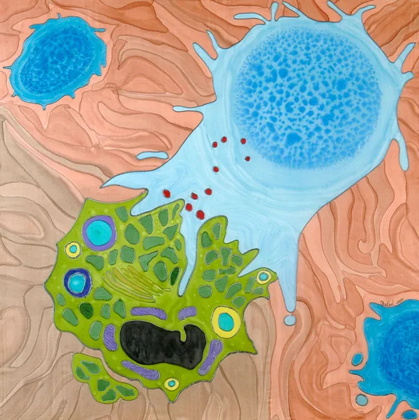
A pale blue immune cell reaching out to a green cancer cell on a background of swirling peach tissue

St George and the Dragon

Odra Noel · paint on silk

Noel trained as a doctor and earned a PhD in basic science before she began painting cells on silk. Here a killer T cell, one of the immune system's defenders, destroys a cancer cell, cast as the knight and the dragon.

CC BY-NC 4.0, Wellcome Collection

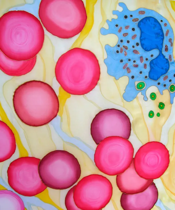
Round magenta red blood cells drifting past a blue white blood cell on a pale yellow background

Blood Cells Through the Microscope

Odra Noel · paint on silk

Part of this painting is what a microscope shows and part is the artist's impression of blood cells and the structures around them.

CC BY-NC 4.0, Wellcome Collection

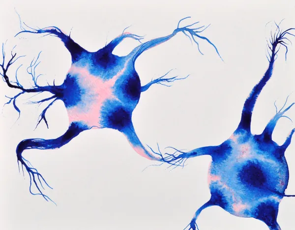
Two deep blue nerve cells with pink centers reaching toward each other with long branching arms on white paper

Neurones Connecting

Stephen Magrath · ink on paper

Neurons are the cells of the nervous system, and they carry information as electrical signals. This painting shows two of them reaching toward each other, the way nerve cells connect to pass a message along.

CC0, Wellcome Collection

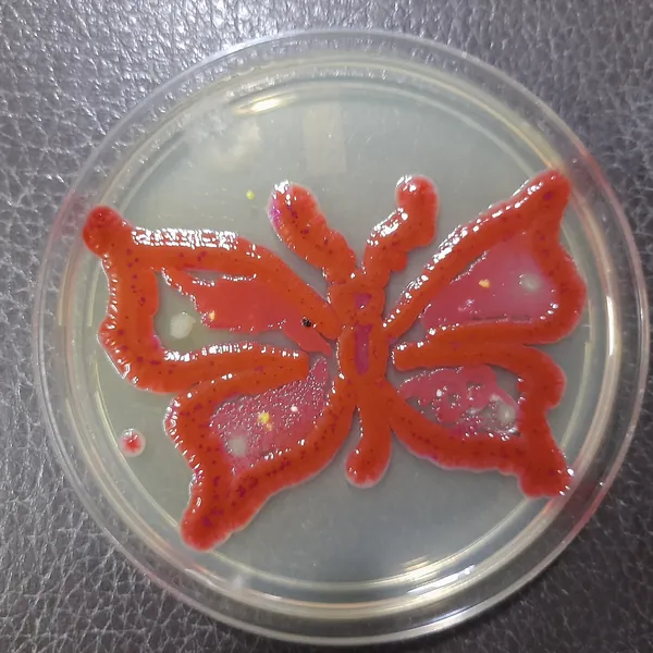
A red butterfly grown from living microbes on the agar in a round glass petri dish

When the petri dish becomes the palette

Microbiologists paint too, with living microbes for paint and a petri dish of agar for a canvas. Ajay Kumar Chaurasiya grew this butterfly in 2021. Alexander Fleming, who discovered penicillin, made germ paintings on agar with colored bacteria, and since 2015 the American Society for Microbiology has run an Agar Art contest. Its 2024 contest drew a record 445 entries.

Sources: American Society for Microbiology, Agar Art contest results, 2024; Hektoen International, Sir Alexander Fleming, 2020.

More artists working in the genre

Their work is protected by copyright, so I describe it here in words. Every one of them is worth looking up.

Klari Reis

Epoxy paintings in petri dishes, San Francisco

Reis pours dyed epoxy into plastic petri dishes and hangs them by the dozen. The work began after she was diagnosed with Crohn's disease and a doctor let her watch her own cells react to different drugs under a microscope.

Michele Banks

Watercolor cells and viruses, Washington, DC

Banks lets wet watercolor bleed into branching patterns that look like blood vessels and brain cells. She has said her best known paintings are her images of mitosis.

Greg Dunn

Neurons in ink and gold, Philadelphia

Dunn earned a doctorate in neuroscience and paints neurons in Japanese sumi-e ink, often on gold leaf. The style began by accident, when he blew on wet ink and it spread into shapes like nerve cells.

Hunter Cole

Drawings in glowing bacteria, North Carolina

Cole, a geneticist, paints with bioluminescent bacteria on petri dishes. The drawings glow blue-green and fade over about two weeks as the bacteria run out of food, so she photographs them while they last.

Luke Jerram

Glass sculptures of microbes, Bristol, England

Jerram, who is color-blind, designs clear glass sculptures of viruses and microbes with virologists. He leaves out color on purpose, to question the bright false colors in many pictures of microbes.

Rogan Brown

Paper sculptures of microbes, France

Brown cuts layered paper into microbes and cells by hand or by laser. His sculpture Outbreak (2014) took about four months to plan, cut and assemble, and he names Ernst Haeckel as an inspiration.
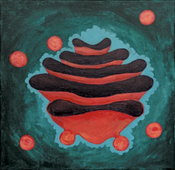
My painting of the Golgi apparatus: stacked dark and red-orange sacs on a teal background, ringed by round vesicles

Where my paintings fit in

I'm Lily Butler, a high school senior, and Petri Palette is my small addition to this tradition. I paint the cell processes high school biology asks students to learn: the stages of mitosis and the path from gene to protein.

Each of my paintings hangs beside a title card that explains the science in plain language. My painting of the Golgi apparatus, shown here, pictures the organelle named for Camillo Golgi, who shared the 1906 Nobel Prize with Cajal.

Seven paintings of the cells biology class teaches

The stages of mitosis and the path from gene to protein, each with a title card I wrote.

Image credits

  1. Robert Hooke, Micrographia (1665), Schem. XI. Scan: National Library of Wales. Public domain, via Wikimedia Commons.
  2. Walther Flemming, Zellsubstanz, Kern und Zelltheilung (1882), Tafel IIIb, cropped from the two-page spread. Wellcome Collection, CC BY 4.0, via Wikimedia Commons.
  3. Santiago Ramón y Cajal, Purkinje neurons, about 1900. Cajal Institute (CSIC), Madrid. Public domain, via Wikimedia Commons.
  4. Illustration by David S. Goodsell, RCSB Protein Data Bank. doi: 10.2210/rcsb_pdb/goodsell-gallery-028. CC BY 4.0.
  5. Illustration by David S. Goodsell, The Scripps Research Institute. doi: 10.2210/rcsb_pdb/goodsell-gallery-011. CC BY 4.0.
  6. Cell fates in zebrafish retina, acrylic painting. Prof. Bill Harris. CC BY 4.0. Source: Wellcome Collection.
  7. St George and the dragon: T lymphocyte killing a cancer cell. Odra Noel. CC BY-NC 4.0. Source: Wellcome Collection.
  8. Blood cells through the microscope. Odra Noel. CC BY-NC 4.0. Source: Wellcome Collection.
  9. Neurones connecting, artwork. Stephen Magrath. CC0 1.0. Source: Wellcome Collection.
  10. Microbial Art, Ajay Kumar Chaurasiya, 2021. CC BY-SA 4.0, via Wikimedia Commons. Cropped; the crop is shared under the same license.
  11. Photo at the top of the page: Pauline Loroy, Unsplash.